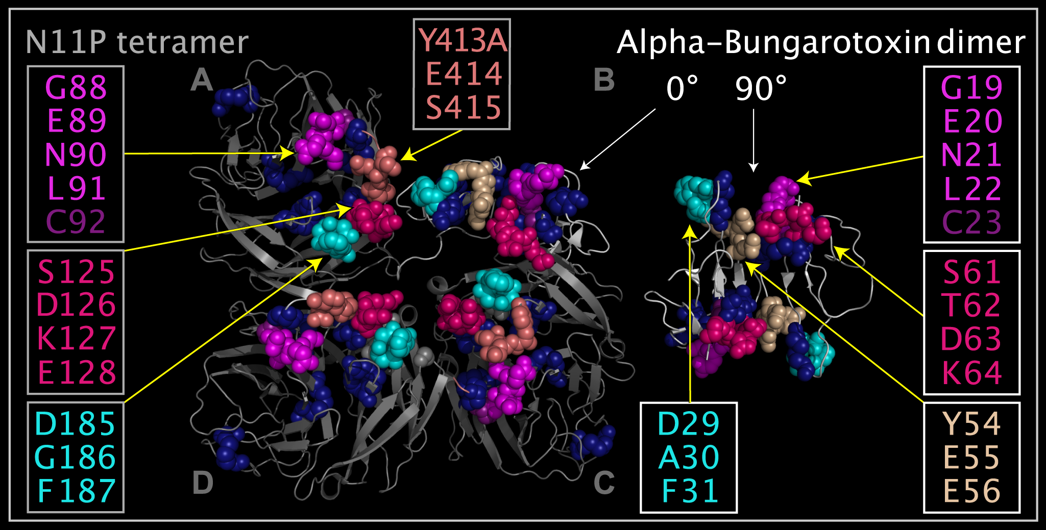
Figure 11. Corresponding residues in alpha-bungarotoxin (“ABT”) dimer and N11 protein (“N11P”) Downside variable loop region (“VLR”).
Shown are two ABT dimers (structure ribbons colored white with dark blue spheres representing disulfide bridges) and a N11P tetramer (structure ribbons colored grey and with dark blue spheres representing disulfide bridges) from the crystal structures. One ABT dimer is shown apart from the N11P tetramer and the other ABT dimer is shown at an angle that is 90 degrees rotated from the first dimer in position “B”, i.e. positioned with the ABT dimer superposed onto and substituting for the N11P monomer in position “B” in the tetramer. Atoms used to superpose the ABT dimer into the position that would be occupied by the N11P “B” monomer are listed in Table 8. N11P Downside VLR residue spheres in monomer positions “A”, “C”, and “D” depicting: G88- L91 colored magenta, C92 colored plum, S125-E128 colored hot pink, D185-F187 colored cyan, and Y413A-S415 colored salmon. Corresponding ABT residue spheres in the two dimers depicting: G19-L22 colored magenta, C23 colored plum, S61-K64 colored hot pink, D29-F31 colored cyan, and Y54-E56 colored tan. N11P monomers in the “A” and “D” positions are shown in the crystal structure positions. In the N11P monomer in the “C” position, the Y413A-S415 residues (colored salmon) have been rotated into the same position relative to G19-L22 (colored magenta) and D29-F31 (colored cyan) as in corresponding residues of ABT crystal structure dimer (shown in the “B” position) in order to illustrate that small movements of the mobile N11P Downside VLR residues can produce nearly identical relative residue presentation to ABT.